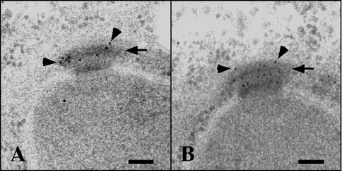
FIG. 2.
Localization of Ady4p-GFP and Spo74p-GFP by electron microscopy. Sporulating cells from strains AN279 (ADY4-GFP/ADY4-GFP) (A) and AN282 (SPO74-GFP/SPO74-GFP) (B) were processed for electron microscopy, and sections were stained with primary antibodies to GFP and gold-conjugated secondary antibodies. Arrows point to MOPs, and arrowheads point to selected gold particles. Bars, 100 nm.

