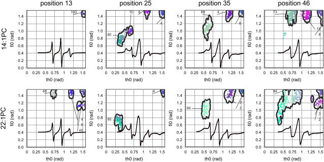
FIGURE 3.
ESR spectra of 3-maleimidoproxyl spin-labeled M13 mutant coat proteins S13C, A25C, A35C, and T46C reconstituted into 14:1 PC and 22:1 PC multilamellar vesicles at L/P 100 in 150 mM NaCl, 10 mM Tris (pH 8.0), and 0.2 mM EDTA at room temperature (black line). Spectral line heights are normalized to the same central line height. Simulated spectra (red lines) were fitted to the experimental ESR spectra (black lines) using an HEO optimization. GHOST condensations plots are given where the parameter ϑ is the cone angle within the spin label can move, and parameter φ describes the asymmetry of the cone. The relative fractions of a group of solutions for the two spectral parameters are indicated for each spin-labeled mutant. The red, green, and blue colors of the solutions codes for the relative values of τc, W, and pA in their definition intervals {0–3 ns}, {0–4 G}, and {0.8–1.2}, respectively (40).

