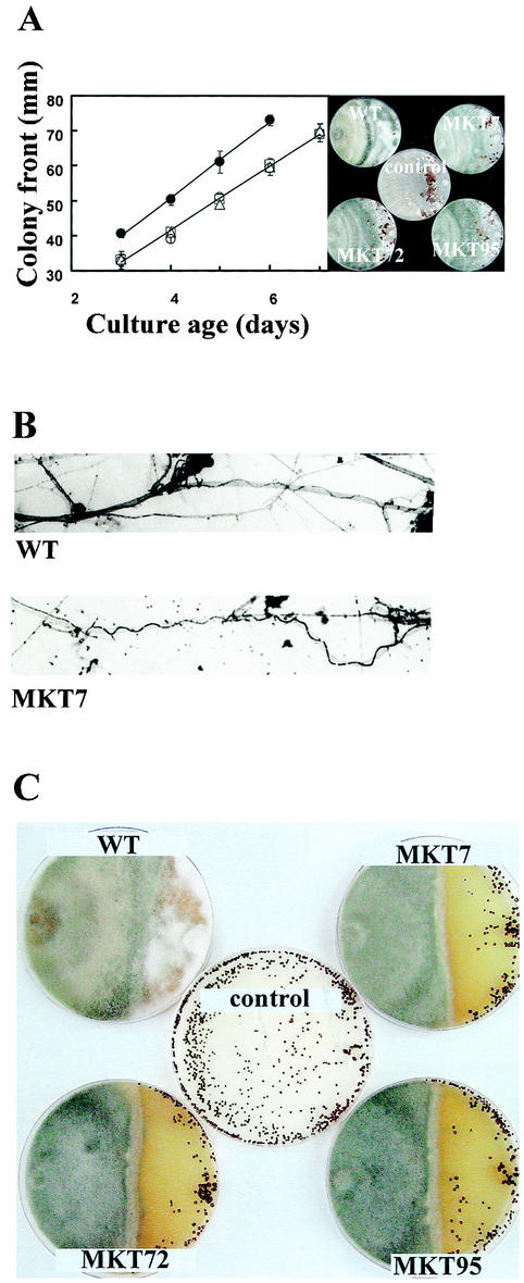
FIG. 5.
Confrontation assays of T. virens and host plant-pathogenic fungi. The host was inoculated at the right side of the plate. Controls, host only. WT, wild type. (A) R. solani versus T. virens. Error bars indicate standard errors for three replicates. Right, typical confrontation plates. Dark structures are the sclerotia of the host. (B) Mycoparasitic coiling of wild-type T. virens and a tmkA mutant on R. solani hyphae 5 days after inoculation. The large-diameter hyphae belong tothe host, while the thinner, dark-stained ones are the mycoparasite, which grows in a wavy or coiling pattern in contact with the host. (C) S. rolfsii versus T. virens, 14 days after inoculation. The dark, roughly spherical structures are the sclerotia of the host. Note that sclerotia are not visible in the wild-type T. virens-S. rolfsii interaction.

