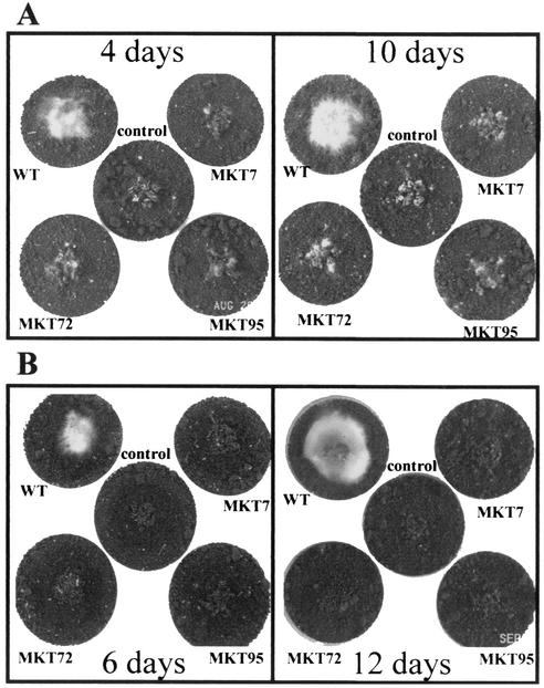
FIG. 6.
Parasitism of sclerotia by T. virens. The sclerotia were put in the middle of a soil plate (see Materials and Methods) after mixing conidia into the soil. Center plates, sclerotia only, with no Trichoderma conidia. Plates were photographed after the indicated number of days of incubation. (A) Growth on R. solani sclerotia. (B) Growth on S. rolfsii sclerotia.

