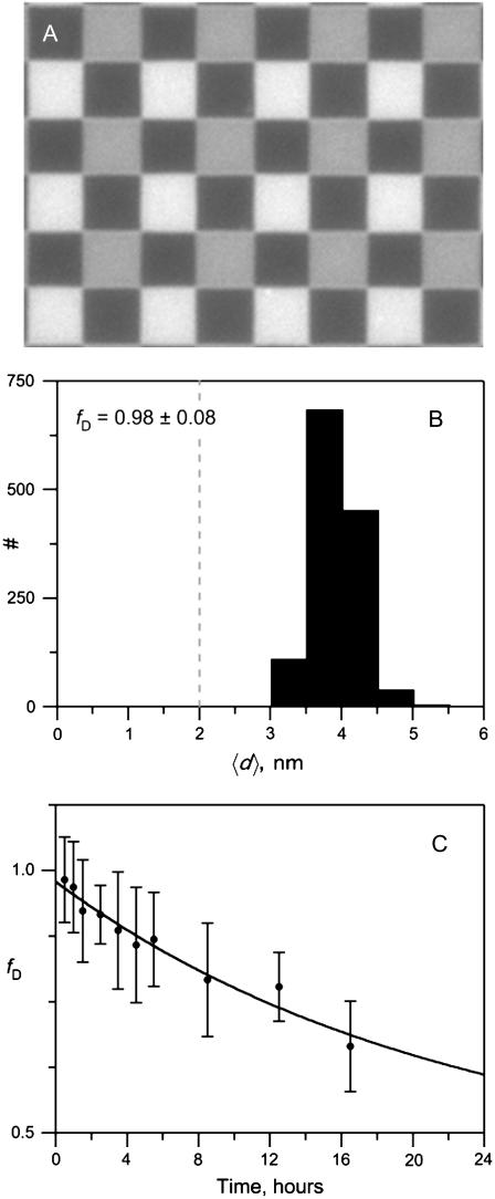
FIGURE 5.
Degradation of lipid asymmetry in planar-supported bilayers monitored by FLIC microscopy. Bilayers were made by the LB/VF technique on 4-oxide FLIC chips at room temperature and stained with 0.5% Rh-DPPE in the vesicles only. (A) Fluorescence micrograph of an asymmetrically stained POPC bilayer on a 4-oxide FLIC chip. Each square on the chip is 5 × 5 μm2. (B) Histogram of measured average distances of dyes from the proximal face of the supported POPC bilayer immediately after its completion. The fraction fD of dye remaining in the distal layer is high at this time. A completely randomized bilayer would have an average dye distance 〈d〉 of 2 nm as indicated by the dashed line. (C) Time course of fraction of Rh-DPPE remaining in the distal monolayer of an asymmetrically stained supported POPC bilayer.

