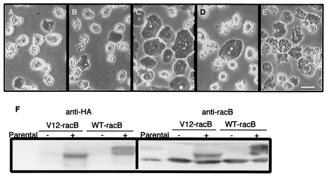
FIG. 1.
Cell morphology changes induced by expression of RacB. (A to E) Cells were grown for 36 h in the presence or absence of folic acid, and phase-contrast images were captured. Wild-type Ax4 cells in the absence of folic acid (A), V12-RacB cells in fresh medium containing 1 mM folic acid (B), and WT-RacB cells (C), V12-RacB cells (D), and WT-RacC cells (E) in the absence of folic acid are shown. The morphology of WT-RacB, V12-RacB, and WT-RacC cells in the presence of folic acid is almost identical to that of parental cells but is dramatically changed on induction. WT-RacB cells became flattened, V12-RacB cells became detached with numerous protrusions on the cell surface, and WT-RacC cells became relatively flat with irregular contours on the surface. Bar, 20 μm. (F) Expression of WT-RacB and V12-RacB. Extracts of 2 × 10⁵ parental, V12-RacB, and WT-RacB cells were harvested and run on an SDS-10% polyacrylamide gel. Expression of RacB was detected by rabbit polyclonal antibody against RacB (right panel) or by anti-HA antibody (left panel). In the presence of folate at low cell density (2 × 10⁵ cells/ml), the recombinant protein was barely detectable (− lanes); however, it was induced twofold over the endogenous RacB level at high cell density in the absence of folic acid (+ lanes).

