FIG. 3.
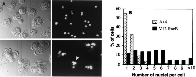
Nuclear staining of parental and V12-RacB cells. (A) Cells were grown for 3 days under induced conditions and stained with 1 μM propidium iodide to visualize nuclei. Left panels are DIC images, and right panels show the nuclear staining. The upper panels are wild-type cells, and the lower panels V12-RacB cells. The arrowhead indicates heterogeneous sizes of nuclei in V12-RacB cells. (B) The number of nuclei was counted from 157 parental and 97 V12-RacB-expressing cells. In parental cells, 87% contain one or two nuclei, as opposed to 26% in V12-RacB population. The number of nuclei in cells expressing V12-RacB ranged from 1 to 15, compared to 1 to 5 in wild-type cells.

