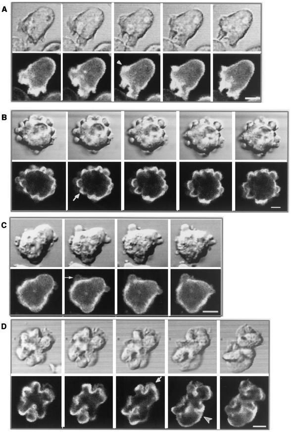
FIG.8.
Visualization of actin dynamics in live cells, using GFP-ABD. (A) Parental Ax4 cells expressing GFP-ABD were observed by confocal microscopy at 20-s intervals. GFP-ABD localizes to the actin cortex and new protrusions as they form at the periphery of the cell (arrow). (B) Cells coexpressing GFP-ABD and V12-RacB were induced for 2 days, and time-lapse images were acquired at 20-s intervals. Cells became detached and actively formed actin-containing spherical protrusions (arrow). (C) Ax4 cells expressing GFP-ABD were electroporated, and the process of bleb formation was visualized by confocal microscopy at 7-s intervals. Note the lack of staining of the bleb. (D) Cells coexpressing GFP-ABD and WT-RacC were induced for 2 days, and time-lapse images were acquired at 20-s intervals. Cells deform the actin cortex both inwardly (arrowhead) and outwardly (arrow). Bar, 5 μm.

