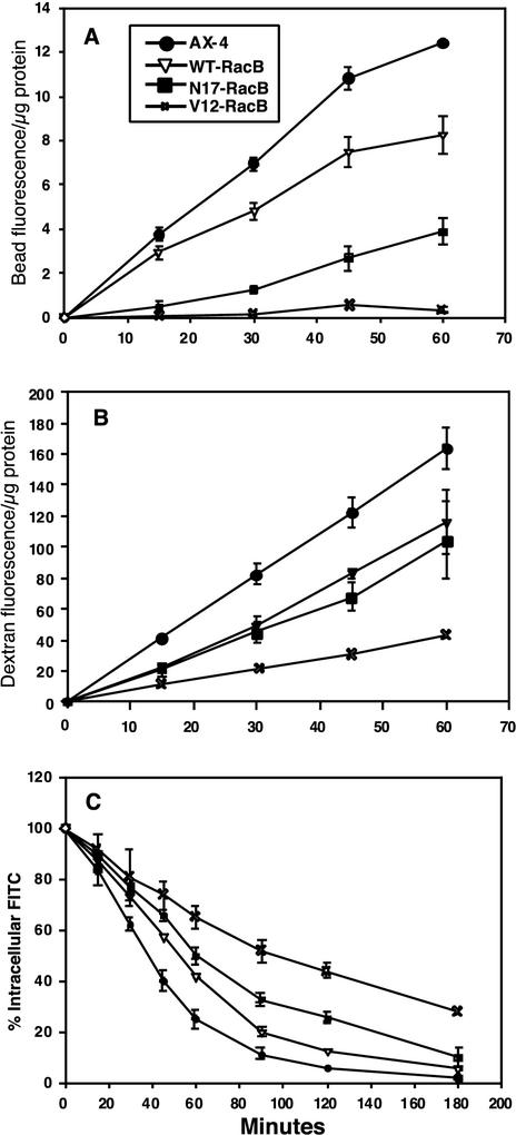
FIG. 9.
Quantitation of phagocytosis, macropinocytosis, and recycling rates. (A) Uptake of fluorescent beads by phagocytosis. (B) Uptake of fluorescent dextran by endocytosis. (C) Efflux of the endocytic probe. Efflux was measured by feeding cells with fluorescent dextran for 3 h and then measuring the fluorescence remaining at each time point.

