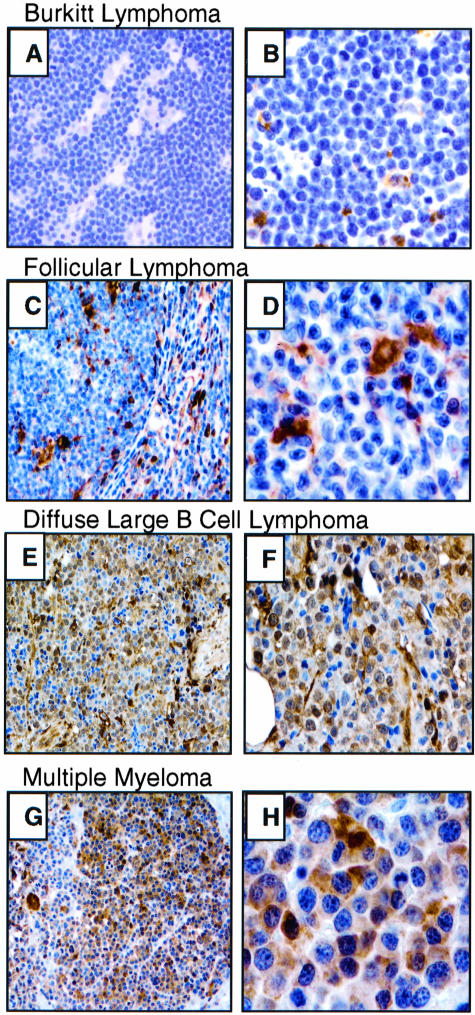
Figure 1.
Galectin-3 immunostaining of representative B-NHL. BL (A and B) and FL (C and D) tumor cells are negative for galectin-3. DLBCL (E and F) and MM (G and H) tumor cells express high levels of galectin-3. Macrophages and dendritic cells, when present within the lymphoma (D), are positive for galectin-3, serving as an internal control for staining.49 Hematoxylin counterstain. Original magnification: A, C, E, G, ×200; B, D, F, ×400; H, ×1000.

