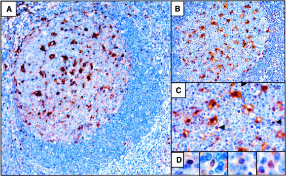
Figure 4.
Galectin-3 expression in tonsil. (A to C) GC follicle showing galectin-3 immunostaining with a hematoxylin counterstain. Positive cells within the GC consist primarily of dendritic reticular cells. The arrows in C indicate scattered, non-dendritic reticular cells staining positive for galectin-3. D: Rare lymphocytes within the GC stain positive for galectin-3. Magnifications: ×100 (A), ×200 (B), ×400 (C), ×600 (D).

