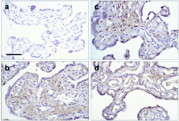
Figure 2.
Immunohistochemistry of TNF-α in tissues sampled immediately after delivery (a), kept under hypoxia (b), normoxia (c), and subjected to H/R (d). There was an increase in the immunostaining, mainly in the stromal area and occasionally in the endothelium, after 7 hours of culture in all conditions as compared to time 0. More staining in the syncytiotrophoblast was also noted after H/R. Bar, 50 μm.

