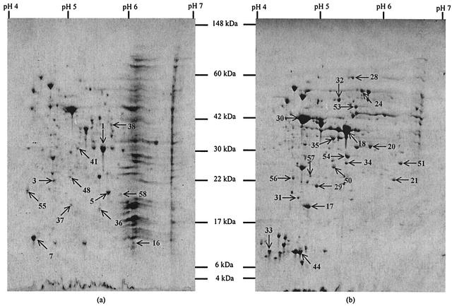
FIG. 3.
Coomassie blue R250-stained 2-D gels of the membrane fraction (a) and cytosolic fraction (b) of protein extracts of L. monocytogenes EGDe. Spots from the two fractions that were excised and identified are numbered. Proteins that were identified are described in Table 1.

