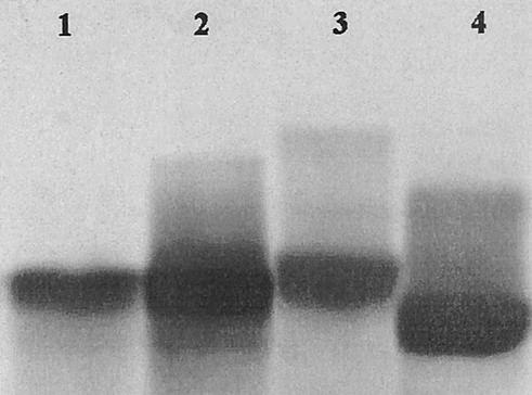
FIG. 1.
Detection of LPS by SDS-PAGE following silver staining. One-microgram samples were analyzed by SDS-PAGE except as noted otherwise. Lanes 1 and 2, M. osloensis at 1 and 5 μg, respectively; lane 3, E. coli EH100; lane 4, E. coli J5. The figure was created with Photoshop 5.5 software (Adobe Systems Inc.).

