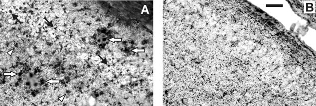
Figure 2.
Representative high-power photomicrographs from the hippocampus of a 17-month-old APP/PS-1 double-transgenic mouse (A) and a 17-month-old control mouse (B), stained with tomato lectin histochemistry for chronically activated microglia. Note the strong activation of microglia in the hippocampus of the APP/PS-1 double-transgenic mouse related to plaques (white arrows) but also within the pyramidal cell layer (CA3, black arrows) and within the molecular layer (arrowheads). Microglial activation was not observed in the hippocampus of 4.5-month-old APP/PS-1 double-transgenic mice or in the hippocampus of the PS-1 single-transgenic mice (data not shown). Bar, 50 μm.

