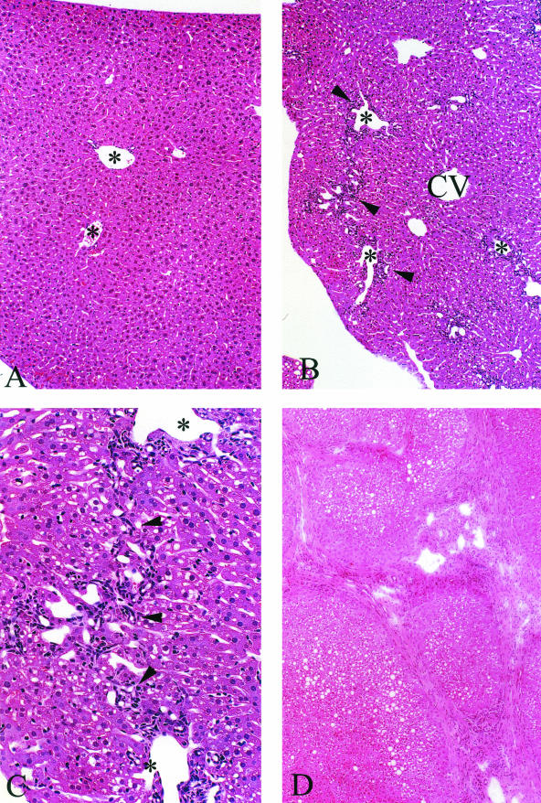
Figure 3.
A: H&E from an 11-month-old wild-type animal maintained on the liquid diet showing no pathological abnormalities. Portal tracts are evident in the field of view (asterisks). B: Cftr−/− littermate shows portal tract inflammation, ductular cell proliferation, and some portal-to-portal bridging (arrowheads). Parenchymal and sinusoidal cells that surround the central vein (CV) appear normal. C: Higher power of B. Portal-to-portal bridging is evident. Arrowheads indicate ductular and inflammatory cells. D: Low power of liver from a 22-month-old Cftr−/− animal that has developed foci of advanced lobular cirrhosis. Original magnifications: ×250 (A, B); ×400 (C); ×200 (D).

