Figure 3.
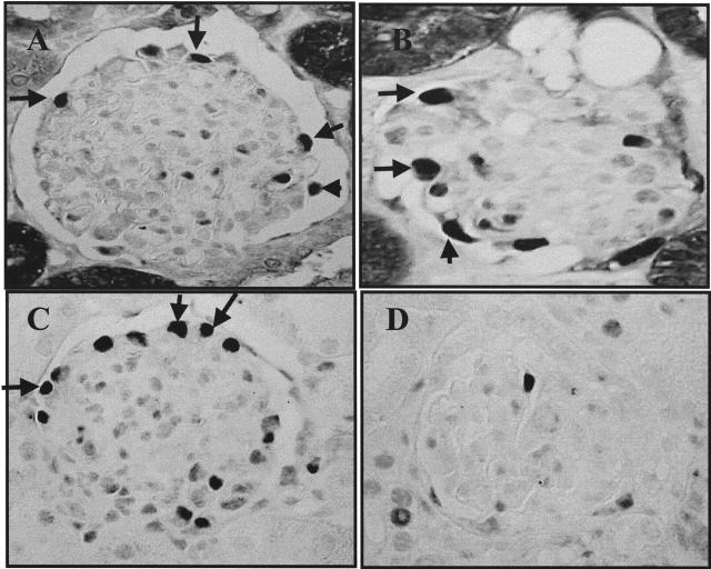
Immunostaining for cyclins D1 and D3 in PHN rats and HIV-transgenic mice. A: Immunostaining for cyclin D1 was increased in PHN rats at day 5, and this was in a typical podocyte localization. B: Cyclin D1 staining increased in HIV-transgenic mice at 6 weeks. C: Cyclin D3 immunostaining was detected in podocytes of PHN rats at day 5, and was unchanged compared to normal. D: In HIV-transgenic mice, podocyte dedifferentiation and proliferation was associated with a decrease in cyclin D3 staining.

