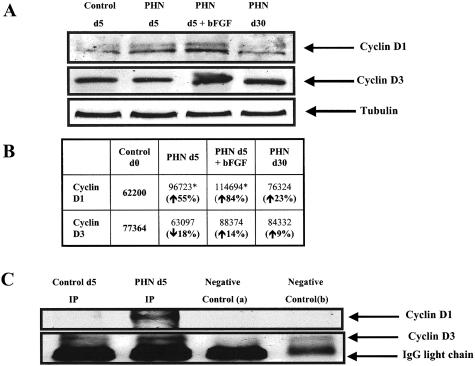
Figure 4.
Western blot analysis for cyclin D1 and cyclin D3 on glomerular protein from PHN rats. A: There was an increase in cyclin D1 protein expression in glomerular protein from day 5 PHN animals (lane 2) compared to control (lane 1), and the increase was more pronounced in PHN rats given bFGF (lane 3). Lane 4 shows that cyclin D1 levels normalized by day 30 PHN. Cyclin D3 protein levels were not changed in PHN. B: Quantification of the Western blots further shows that cyclin D1 levels increase in PHN, however, cyclin D3 levels did not change significantly. C: Co-immunoprecipitating glomerular protein from PHN day 5 rats with an antibody to cdk4 showed a positive signal for cyclin D1 (lane 2), which was absent in glomerular protein from control rats (lane 1).

