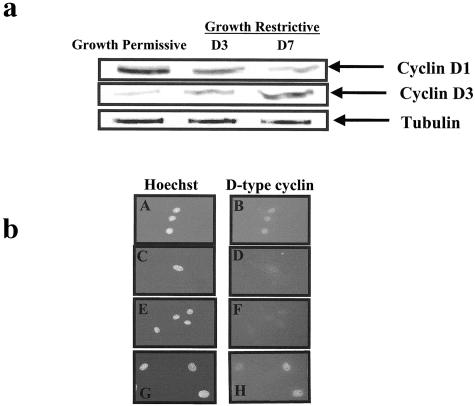
Figure 5.
A: Western blot analysis for cyclin D1 and cyclin D3 in conditionally immortalized podocytes in vitro. Cyclin D1 was abundant in proliferating podocytes grown under permissive conditions. The levels of cyclin D1 decreased when podocytes were grown under restrictive conditions, and this coincided with a decrease in proliferation, and the development of a differentiated phenotype. There was a progressive increase in cyclin D3 levels when grown under restrictive conditions, compared to permissive conditions. Tubulin was used to assure equal loading. b: Immunofluorescence for cyclin D1 and cyclin D3 in conditionally immortalized podocytes. Cyclin D1 stains positive in proliferating podocytes grown under permissive conditions (b), and is barely detected under growth restrictive conditions (D). Hoechst staining was used as a nuclear counterstain (A and C). Cyclin D3 staining was not detected in proliferating podocytes (F), but was abundant in quiescent and differentiated podocytes grown under growth restrictive conditions (H). Hoechst staining was used as a nuclear counterstain (E, G).

