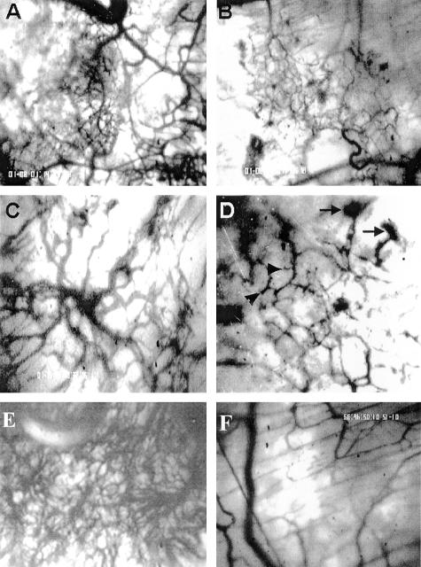
Figure 2.
Effect of ATE + 31 on MCF-7 tumor angiogenesis. Spheroids containing MCF-7 cells were placed within dorsal skin-fold chambers and locally treated (superfused) with 20 ng of ATE + 31 in 20 μl saline or an identical volume of saline alone at 24 hours and every 2 days after. Vascular development was followed by intravital microscopy. A–D: Angiogenesis after 5 days. A: Control, showing robust vascular infiltration. B: ATE + 31-treated, demonstrating little neovascularization. Magnification, ×40. C and D: The integrity of the newly formed blood vessels appears to be aberrant in ATE + 31-treated tumors. Vessels within treated tumors (D) appear fractured and in certain cases end precipitously (arrowheads) or in a mass of poorly formed vessels (arrows) compared to vessels within untreated tumors (C). Magnification, ×100. E and F: Angiogenesis after 15 days. The untreated tumor (E) has a much richer vascular network than the ATE + 31-treated tumor (F). Magnification, ×100.

