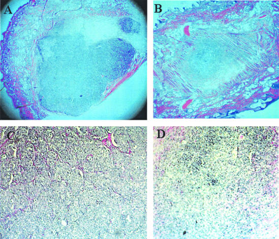
Figure 3.
Histological evaluation of MCF-7 tumors after ATE + 31 treatment. Tissue containing the tumor spheroid, with and without treatment with ATE + 31, was excised after 15 days, fixed, sectioned, and stained with H&E. A comparison of control (A) and ATE + 31-treated (B) tumors shows a dramatic difference in tumor size. Magnification, ×25. A section from untreated tumors under higher magnification (C) shows an abundant vascular supply (stained red) infiltrating the tumor from the surrounding tissue while few vessels can be seen in the section from the ATE + 31-treated tumors (D). Magnification, ×100.

