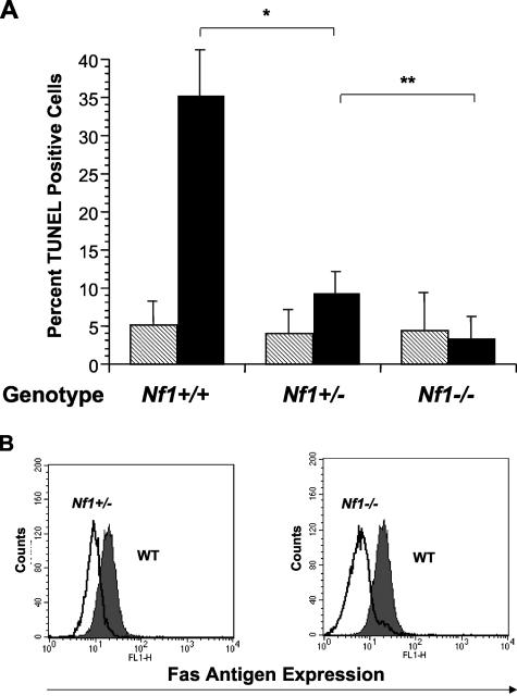
Figure 1.
A–B: Neurofibromin-deficient mast cells are resistant to Fas ligand-mediated apoptosis and have reduced surface Fas antigen expression. A: WT, Nf1 +/−, or Nf1 −/− mast cells were cultured in serum-enriched medium and kit-L for 72 hours. Following culture, cells were stimulated with either Fas ligand (black bars) or vehicle (hatched bars). The percentage of cells undergoing apoptosis was determined by FACS analysis using the TUNEL method. Results represent the mean ± SEM of five independent experiments. * P < 0.03 for comparison of Fas ligand-treated versus vehicle-treated mast cell cultures within each experimental genotype by Student’s paired t-test. ** P < 0.05 for comparison of Fas ligand-treated Nf1 +/− mast cells versus Fas ligand-treated Nf1 −/− mast cells by Student’s paired t-test. B: WT, Nf1 +/−, or Nf1 −/− mast cells were cultured in serum-enriched medium and kit-L for 72 hours and analyzed for expression of surface Fas antigen following culture. The dark profile represents Fas antigen expression by WT mast cells and the overlays represent Fas antigen expression by Nf1 +/− and Nf1 −/− cells in the left and right panels, respectively. Data are representative of five other independent experiments with similar results.

