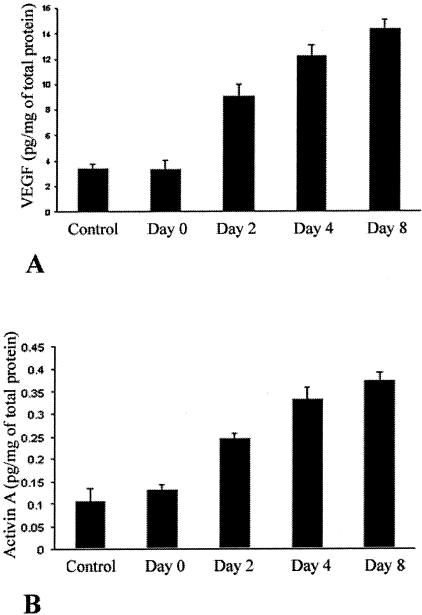
Figure 3.
Protein levels of activin A increase systemically during the course of corneal neovascularization in our murine model and correlate with the increases in the VEGF protein levels. Mice were treated as described in the Materials and Methods section, sacrificed at various days after the scraping, and the levels of activin A and VEGF were measured in corneal lysates. Bars represent the VEGF levels (A) and the activin A levels (B) of the corneal lysates at the indicated days after the corneal scraping (mean ± SD). As previously described, endogenous VEGF protein levels increase steadily after scraping (D0), on days 2 (D2), 4 (D4), and 8 (D8) (A). In parallel, endogenous levels of activin A increase also after the corneal scraping on days D2, D4, and D8, correlating with the increases in endogenous VEGF levels.

