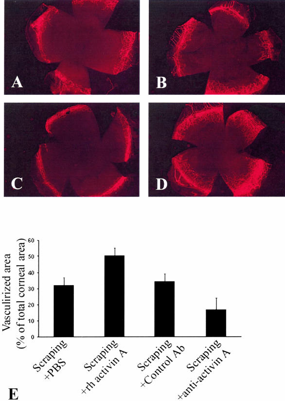
Figure 6.
Activin A regulates corneal neovascularization in the scrape murine model. A to D: Mice were sacrificed on day 12 (D12) after the scraping and corneal vasculature was labeled with FITC-labeled ConA as described in the Materials and Methods section. Representative microscopic images of scraped murine corneas are shown: A: Scraped and treated with vehicle; B: scraped and treated with the isotype-matched control antibody; C: scraped and treated with the neutralizing antibody against activin A; or D: scraped and treated with activin A. E: The vascularized corneal surface expressed as a percentage of the total corneal area was calculated as described in the Materials and Methods section for the various groups (mice scraped and received the vehicle, mice scraped and received activin A, the neutralizing antibody against activin A, or the isotype-matched control). Given that unscraped murine corneas are not vascularized (therefore, percentage of vascularized corneal area ∼0%), scraped murine corneas show increased vascularization and administration of recombinant activin A increases even further the amount of corneal neovascularization, whereas neutralization of endogenous activin A decreases the amount of corneal neovascularization.

