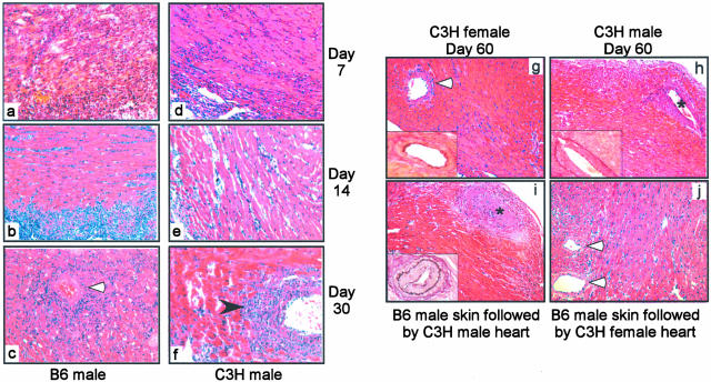
Figure 5.
B6 male heart grafts and C3H male heart grafts transplanted in Mar females exhibit different pathological features. a to f: H&E-stained sections of cardiac allografts obtained 7 (a, d), 14 (b, e), and 30 (c, f) days after transplantation of B6 male (a–c) or C3H male (d–f) hearts. Note the diffuse mononuclear infiltrate in the B6 male hearts and the perivascular infiltrate particularly prominent in C3H male hearts on day 30 (black arrowhead). The photographs are fully representative of three to four animals evaluated per group. g to i: H&E-stained sections with insets showing elastin staining of C3H male or female heart grafts 60 to 70 days after transplantation in Mar females. C3H female hearts transplanted in Mar females (g) exhibited normal histology without evidence of vasculopathy. C3H male hearts transplanted into naïve Mar females (h) or into Mar females that previously rejected B6 male skin (i) developed classic transplant vasculopathy. j: H&E-stained female C3H heart graft in a Mar female harvested 40 days after rejection of a B6 male skin graft. Note the normal histological appearance and absence of infiltrates or vasculopathy. The photographs are fully representative of three to four animals evaluated per group. White arrowhead, blood vessels; *, blood vessels with vasculopathy. Original magnifications: ×40 (a–f and insets); ×20 (g–i).

