Figure 6.
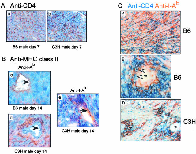
Antigen expression patterns differ in B6 male versus C3H male heart grafts transplanted into Mar female recipients. A: Recipient CD4 T cells traffic to both B6 male (a) and C3H male (b) heart grafts. Frozen graft sections were obtained on day 7 after transplant and stained with anti-CD4 mAb. The results are fully representative of three to four animals studied per group. Quantitative image analysis using ImagePro software revealed no difference in the amount of CD4 detected between the two groups (P < 0.05, data not shown). B: Single-color immunohistochemical staining for I-Ab (c and d) or I-Ak (e) in heart grafts obtained on day 14 after transplant. Note that I-Ab is expressed on the vascular endothelium (black arrowheads) of the B6 grafts, but in the perivascular area (and not on the endothelium) of the C3H grafts. A representative vessel from a C3H graft stained with anti-I-Ak is shown in e as a specificity control. Each photograph is fully representative of three individual animals studied per group. Similar expression patterns were detected at 3 weeks after transplant (not shown). C: Two-color immunohistochemistry for I-Ab (orange) and CD4 (blue) of a B6 male heart graft (f and g) and a C3H male heart graft (h) obtained on day 21 after transplant. Note that in the B6 heart grafts there is diffuse staining for both I-Ab and CD4 (f) and a close association between CD4 T cells and I-Ab-expressing endothelial cells of a blood vessel (white arrowheads, g). In contrast, C3H heart grafts show co-localization of CD4 and I-Ab only in the perivascular area (h). The findings are fully representative of three to four animals studied per group. *, Blood vessel. Original magnifications: ×20 (a–b, f, h); ×40 (c–e, g).

