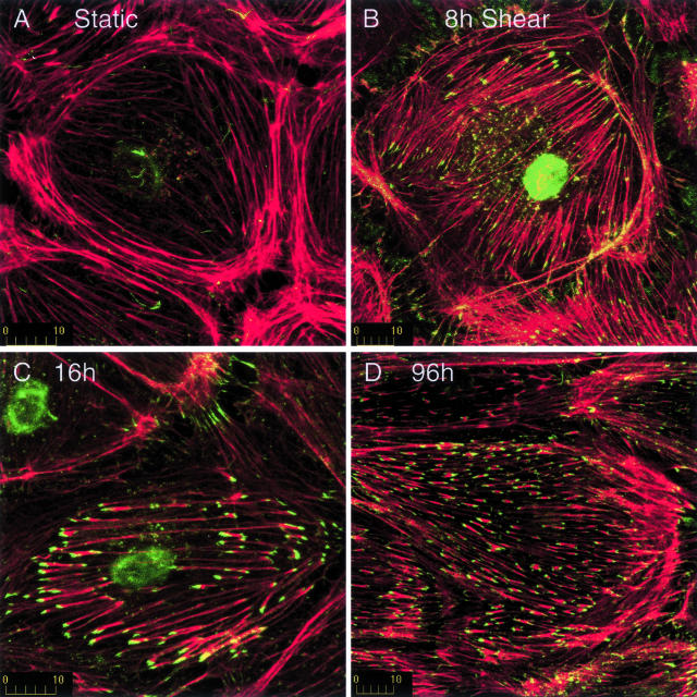
Figure 3.
Shear stress induces assembly of actin at the ends of stress fibers. Endothelial cells were incubated briefly with fluorescently labeled G-actin (green) after exposure to shear stress for 0 hours (A), 8 hours (B), 16 hours (C), or 96 hours (D). Cells were then fixed and filamentous actin was stained with rhodamine phalloidin (red). Little or no actin assembly occurred in postconfluent cells not exposed to shear stress (A). In contrast, exposure to shear stress induced actin assembly at the ends of stress fibers (B–D). Initially, actin assembly was randomly oriented, but assembly became aligned with shear stress by 16 to 24 hours. Shear stress is directed from right to left. Each finding is representative of at least three replicate experiments.

