Abstract
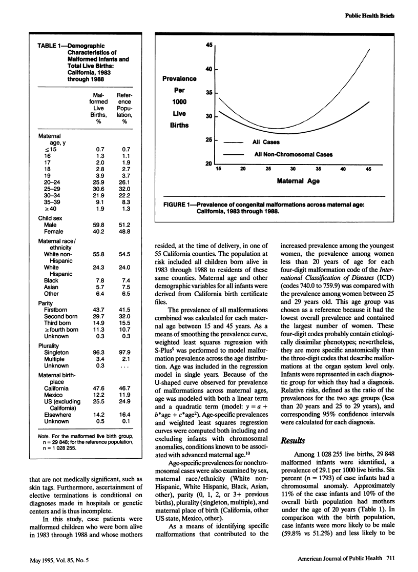
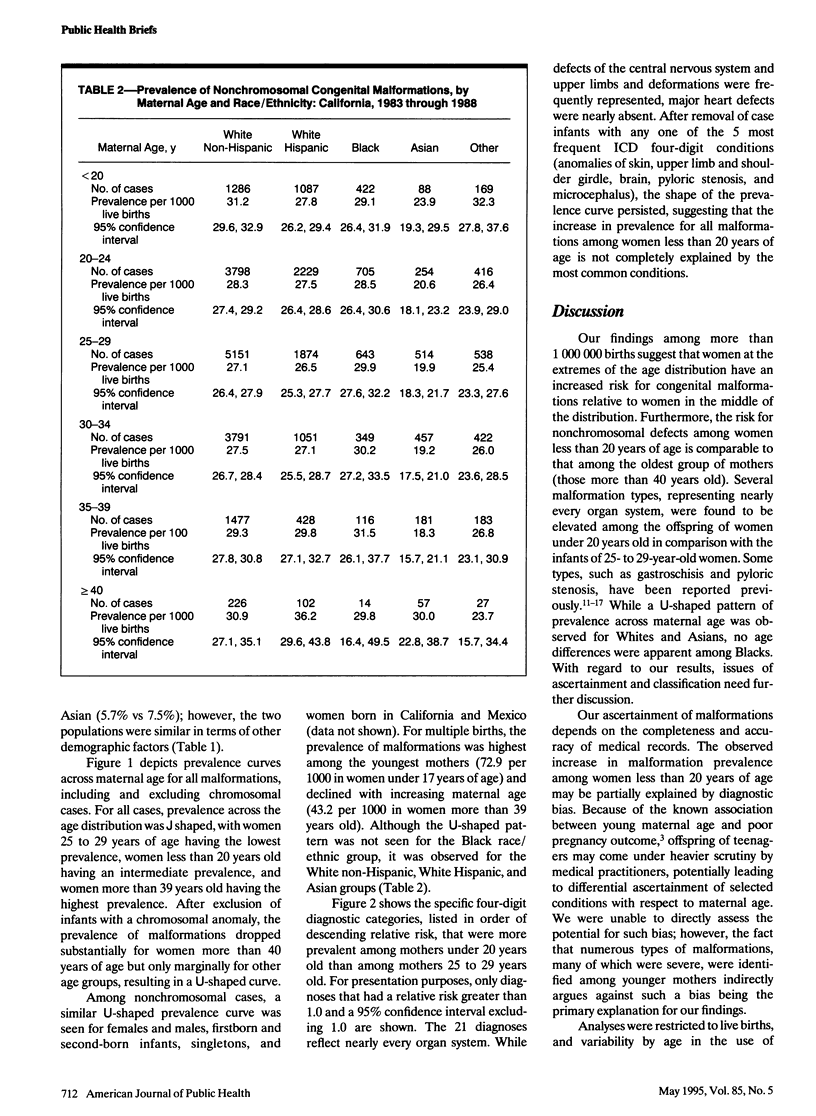
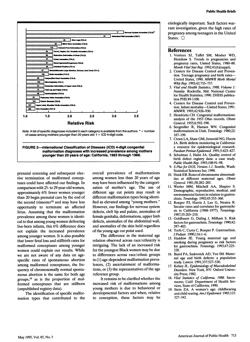
This study examined the prevalence of congenital malformations across the maternal age spectrum and identified specific malformation types that contributed to the overall prevalence among mothers under the age of 20 years. Data were derived from the California Birth Defects Monitoring Program for 1983 through 1988 live births. The distribution of prevalences of all nonchromosomal malformations was U-shaped across maternal age. Furthermore, several specific malformation types, representing nearly every organ system, were elevated among the infants of women under 20 years of age in comparison with those of women 25 to 29 years old.
Full text
PDF


Selected References
These references are in PubMed. This may not be the complete list of references from this article.
- Baird P. A., Sadovnick A. D., Yee I. M. Maternal age and birth defects: a population study. Lancet. 1991 Mar 2;337(8740):527–530. doi: 10.1016/0140-6736(91)91306-f. [DOI] [PubMed] [Google Scholar]
- Croen L. A., Shaw G. M., Jensvold N. G., Harris J. A. Birth defects monitoring in California: a resource for epidemiological research. Paediatr Perinat Epidemiol. 1991 Oct;5(4):423–427. doi: 10.1111/j.1365-3016.1991.tb00728.x. [DOI] [PubMed] [Google Scholar]
- Goldbaum G., Daling J., Milham S. Risk factors for gastroschisis. Teratology. 1990 Oct;42(4):397–403. doi: 10.1002/tera.1420420408. [DOI] [PubMed] [Google Scholar]
- HENDRICKS C. H. Congenital malformations; analysis of the 1953 Ohio records. Obstet Gynecol. 1955 Dec;6(6):592–598. [PubMed] [Google Scholar]
- Haddow J. E., Palomaki G. E., Holman M. S. Young maternal age and smoking during pregnancy as risk factors for gastroschisis. Teratology. 1993 Mar;47(3):225–228. doi: 10.1002/tera.1420470306. [DOI] [PubMed] [Google Scholar]
- Hook E. B. Rates of chromosome abnormalities at different maternal ages. Obstet Gynecol. 1981 Sep;58(3):282–285. [PubMed] [Google Scholar]
- Roeper P. J., Harris J., Lee G., Neutra R. Secular rates and correlates for gastroschisis in California (1968-1977). Teratology. 1987 Apr;35(2):203–210. doi: 10.1002/tera.1420350206. [DOI] [PubMed] [Google Scholar]
- Schulman J., Hahn J. A. Quality control of birth defect registry data: a case study. Public Health Rep. 1993 Jan-Feb;108(1):91–98. [PMC free article] [PubMed] [Google Scholar]
- Seegmiller R. E., Hansen W. N. Congenital malformations in Utah. Teratology. 1980 Oct;22(2):187–199. doi: 10.1002/tera.1420220208. [DOI] [PubMed] [Google Scholar]
- Stein Z. A. A woman's age: childbearing and child rearing. Am J Epidemiol. 1985 Mar;121(3):327–342. doi: 10.1093/oxfordjournals.aje.a114004. [DOI] [PubMed] [Google Scholar]
- Torfs C., Curry C., Roeper P. Gastroschisis. J Pediatr. 1990 Jan;116(1):1–6. doi: 10.1016/s0022-3476(05)81637-3. [DOI] [PubMed] [Google Scholar]
- Werler M. M., Mitchell A. A., Shapiro S. Demographic, reproductive, medical, and environmental factors in relation to gastroschisis. Teratology. 1992 Apr;45(4):353–360. doi: 10.1002/tera.1420450406. [DOI] [PubMed] [Google Scholar]


