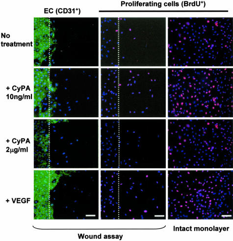
Figure 2.
CyPA stimulates endothelial wound healing (left and middle rows) and proliferation in vitro. Scratch wound assay (dotted line indicates wound edge) showed that the low CyPA concentration tested (10 ng/ml) enhanced migration of ECs (detected with anti-CD31, green fluorescence). Nuclei are counterstained with Hoechst (blue). ECs migrating past the wound edge were quantified after 24 hours. Middle and right rows: EC proliferation, detected using incorporation of BrdU (red), in subconfluent wounded or intact monolayers under different conditions (immunofluorescence). Nuclei of proliferating cells appear as pink. CyPA (10 ng/ml) increased basal level (no treatment) of cell proliferation to a level comparable to that induced by VEGF, used as a positive control. A biphasic effect was observed, with high CyPA concentrations (2 mg/ml) decreasing cell proliferation.

