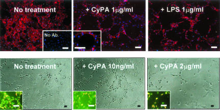
Figure 4.
In vitro endothelial tube formation assay. Top: ECs highlighted by immunofluorescence using anti-CD31 (red), nuclei counterstained with Hoechst (blue). LPS and CyPA treatments of HUVECs appear to have similar capacity to induce formation of EC tubes on Matrigel. Right bottom inset in no treatment section illustrates the negative control for immunocytochemistry (no Ab, no primary antibody). Left bottom inset in CyPA 1 mg/ml section illustrates at higher magnification the appearance of the lumen of a tube. Bottom (phase contrast): tube formation assay performed on growth factor-reduced Matrigel. The total number of tubes was increased ∼10.5-fold at 10 ng/ml of CyPA compared with the nontreated control (P = 0.0021), but decreased at high concentrations (2 μg/ml of CyPA). Insets (live-dead assay) illustrate effects on cell viability likely contributing to the differences—note the increased percentage of dead cells (red fluorescence) compared to live cells (green). Scale bars, 50 μm.

