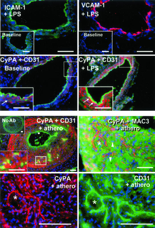
Figure 6.
In vivo vascular expression of CyPA is associated with acute and chronic pathological conditions. Immunofluorescence analysis of mouse carotid artery cross-sections from healthy, untreated mice (baseline), mice injected with LPS (+LPS), or from mice with experimental atherosclerotic lesions (+ athero). Blue fluorescence, cell nuclei counterstained with Hoechst. Top: LPS injection induces acute endothelial activation, as indicated by induction of ICAM (green fluorescence) and VCAM-1 (red fluorescence) expression. Compare to nondetectable baseline expression (insets) of these molecules in normal endothelium. Second row: Luminal endothelial layer is detected using anti-C31 (green fluorescence). Low baseline (left) level of CyPA expression (red fluorescence) is detectable within the normal arterial wall in smooth muscle cells (inset, arrows) but not in luminal ECs. Right: Injection of LPS (+LPS) increased expression of CyPA throughout the wall and induced expression associated with the luminal endothelium (yellow fluorescence). Bottom left inset illustrates the higher magnification of the boxed area. Note the appearance of the yellow signal (arrows), indicating co-localization of CyPA (red) and luminal ECs (green). Bottom: Experimental atherosclerotic lesions induced in the carotid artery of ApoE−/− mice (+athero) a model for a chronic pathological condition of arteries. Top: CyPA (red) is detected diffusely within the intimal lesion and the medial layer. Left: CyPA did not appear associated with the luminal endothelium (thick arrow) but was co-localized (small arrows) with the ECs (green) of neocapillaries that develop within these lesions. Bottom left inset is a higher magnification of the boxed area illustrating the yellow signal in capillaries. Top inset illustrates the negative control for immunohistochemistry, consecutive section processed in the absence of primary antibodies (no Ab). Right: Simultaneous detection of CyPA (red) and macrophages (green), using anti-MAC3, suggesting little overlap. Note some obviously negative macrophages (arrowheads). Bottom: At higher magnification the staining patterns of consecutive sections (the asterisk indicates the same tissue area) for CyPA (red) and endothelium (CD31, green). Although CyPA expression is not restricted to the neocapillaries, these are clearly positive for CyPA. Scale bars, 50 μm.

