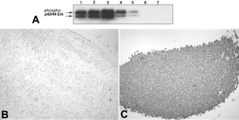
Figure 1.
Calibration of phospho-ERK immunohistochemistry. A: Western blot for phospho-ERK in whole cell lysates of SKBR3 cells under different conditions: lane 1, 10% fetal bovine serum (FBS); lane 2, 10% FBS and 10 ng/ml EGF; lane 3, 10% FBS and 20-minute exposure to EGF; lane 4, no serum; lane 5, no serum and 10 μmol/L MEK inhibitor UO126 (Promega); lane 6, no serum and 25 μmol/L UO126; lane 7, no serum and 75 μmol/L UO126. B: Negative control: SKBR3 cells were grown in the absence of serum and 10 μmol/L MEK inhibitor UO126 (Promega), formalin-fixed, paraffin-embedded, and stained with anti-phospho-ERK-1/2 antibody at 1:200. C: Positive control: SKBR3 cells were grown in 10% FBS and 20-minute exposure to EGF, formalin-fixed, paraffin-embedded, and stained with anti-phospho-ERK-1/2 antibody at 1:200.

