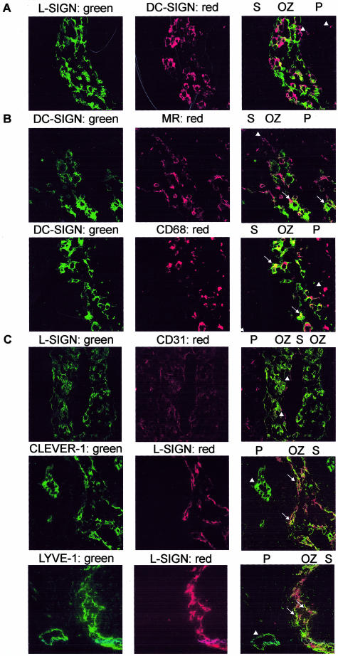
Figure 3.
In the cortical outer zone, DC-SIGN+ cells express mannose receptor and CD68 intracellularly and L-SIGN+ cells co-express LYVE-1 and CLEVER-1. A: Tissues sections of lymph nodes (patient 1) double stained with AZN-D1 and PTTS, followed by anti-mouse Texas Red and anti-rabbit FITC, and analyzed by confocal microscopy. Arrowheads indicate DC-SIGN+L-SIGN− cells in the paracortex. B: Sections (patient 1) were double stained with CSRD, followed by anti-rabbit FITC and anti-CD68 or anti-mannose receptor, followed by anti-mouse Texas Red. C: Sections (patient 1) were double stained with PTTS, followed by anti-rabbit FITC and anti-CD31 and by anti-mouse Texas Red or with PTTS and anti-rabbit Texas Red in combination with anti-LYVE-1 or anti-CLEVER-1 and anti-mouse Alexa 488. Arrowheads indicate single-positive cells (CD68+, mannose receptor+, LYVE-1+, or CLEVER-1+), arrows point to double-positive cells. S, sinus; OZ, outer zone of paracortex; P, paracortex. Original magnifications, ×40.

