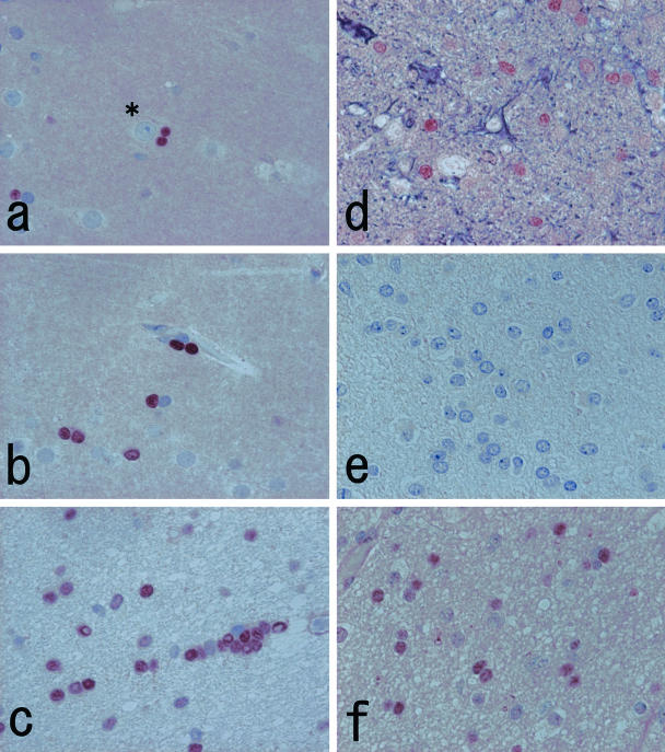
Figure 3.
a to c: Immunohistochemistry with Olig2-C labels nuclei of the cells of perineuronal (a), perivascular (b), and interfascicular (c) dispositions. The size of immunolabeled nuclei is smaller than that of neurons (asterisk) and astrocytes. These characteristics are compatible with oligodendrocytes. d: Double immunostaining of Olig2-C (brown) and anti-glial fibrillary acidic protein antiserum (deep purple) demonstrates a nonoverlapping positive reaction. e: The nuclear staining is remarkably reduced when the primary antibody is replaced with a preabsorbed fraction of Olig2-C. f: Immunohistochemistry with Olig2-N displays weaker nuclear staining with diffuse background noise. Original magnifications, ×500 (a–f).

