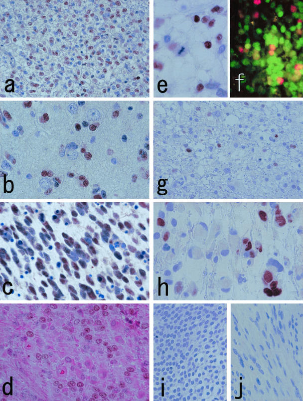
Figure 4.
a and b: Oligodendroglioma cells with perinuclear haloes (a) and those showing perineuronal satellitosis (b) are stained by Olig2-C. Note that not all nuclei are immunostained. c: Olig2-C reaction is noted in anaplastic oligodendroglioma. This specimen is highly mitotic and accompanied by abundant apoptotic bodies. The apoptotic nuclei are Olig2-C-negative. d: Double staining of Olig2-C and H&E on anaplastic oligodendroglioma illustrates that cells with astrocytic differentiation are weakly or not stained by Olig2-C. e: A nucleus under mitotic phase is Olig2-C-negative (anaplastic oligodendroglioma). f: Dual-immunofluorescent staining with MIB-1 (red) and Olig2-C (green) illustrates MIB-1-positive/Olig2-C-negative cells in addition to some double-positive cells (anaplastic oligodendroglioma). g and h: Representative cases of astrocytic tumors (g, fibrillary astrocytoma; h, glioblastoma). Scattered Olig2-C-positive nuclei are noted. The cell bodies possessing labeled nuclei appear vacant or inconspicuous, whereas truly astrocytic cells with rich cytoplasm tend to be Olig2-C-negative. i and j: Central neurocytoma (i) and schwannoma (j) are negative with Olig2-C. Original magnifications: ×250 (a, f, g, i, j); ×500 (b–e, h).

