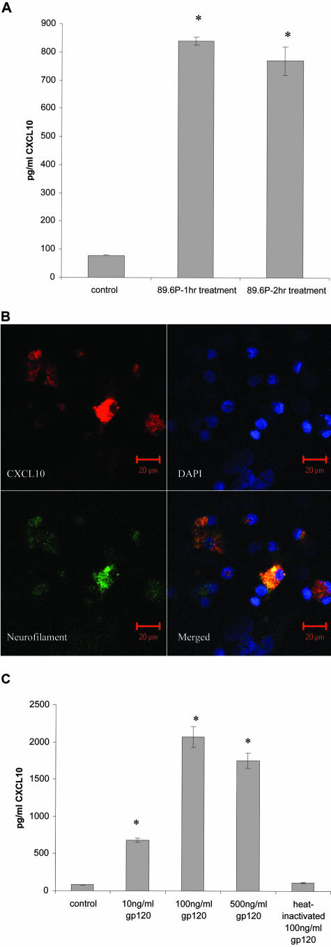
Figure 3.
Fetal neuronal cultures were exposed to SHIV89.6P (A) or varying concentrations of macrophage-tropic gp120BAL (C) and expression of CXCL10 in the culture supernatants was quantitatively determined by ELISA. *, Statistical significance compared to control. B: SHIV89.6P induced CXCL10 expression in neurons in fetal brain cultures. Fetal neuronal cultures treated with the virus stock were immunostained with anti-CXCL10 (red) and anti-neurofilament (green) antibodies. DAPI (blue stain) was also applied to stain the nuclei. CXCL10 was localized in neurons.

