Figure 1.
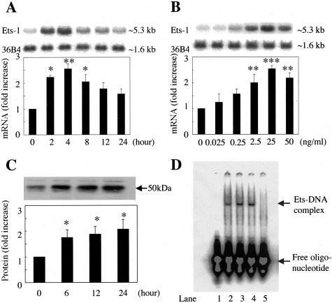
Stimulation of Ets-1 expression by VEGF. A: Time-dependent induction of Ets-1 mRNA after stimulation by VEGF. Total RNA was isolated at the indicated time points after the cells were stimulated with VEGF (25 ng/ml), and Northern blot analysis was performed. B: Dose-dependent induction of Ets-1 mRNA 4 hours after stimulation by VEGF. BRECs were treated with the indicated concentrations of VEGF for 4 hours. Representative Northern blots and control 36B4 (top) and quantitation after normalization to the control signals are shown (bottom). C: Effect of VEGF on Ets-1 protein expression. BRECs were studied after 6, 12, and 24 hours of incubation with VEGF (25 ng/ml). Total protein (30 μg) was assessed by Western blot analysis using anti-Ets-1 antibody. The size of Ets-1 protein corresponded to ∼55 kd. Representative Western blots (top) and quantitation (bottom). Results are shown as fold increases of control. Mean ± SEM of three separate experiments (each in triplicate). *, P < 0.05; **, P < 0.01; ***, P < 0.001. D: Stimulation of DNA-binding activity by VEGF. DNA-binding activity of nuclear protein was analyzed by EMSA. BRECs were incubated for 12 or 24 hours with VEGF (25 ng/ml), and then nuclear protein was obtained and applied to EMSA. Lane 1, negative control (free probe alone); lane 2, BRECs basal level (control); lane 3, VEGF stimulation for 12 hours; lane 4, VEGF stimulation for 24 hours; lane 5, VEGF stimulation for 12 hours with unlabeled competitor.

