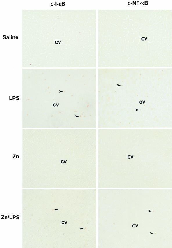
Figure 4.
In situ detection of LPS-induced NF-κB activation in the liver by immunohistochemistry. MT-KO mice were administrated with one dose of LPS at 4 mg/kg. Livers were removed at 1 hour after LPS challenge, and cryostat sections were cut at 5 μm. NF-κB activation was monitored by immunohistochemical staining of phospho-I-κB (p-I-κB) and phospho-NF-κB (p-NF-κB). Arrowheads show positive cells immunoreactive to p-I-κB and p-NF-κB. CV, Central vein. Original magnifications, ×260.

