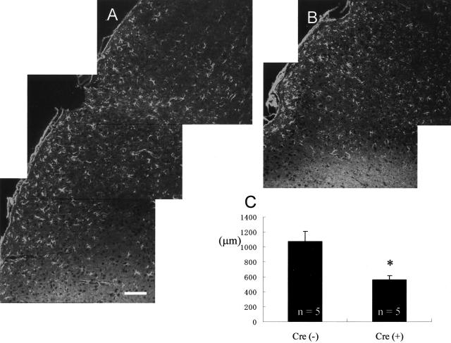
Figure 4.
The astrogliosis, detected by an increased level of GFAP immunoreactivity in astrocytes, was enhanced in the brain of Cre(−) compared to Cre(+) mice (A and B, respectively). The average width of the astrogliosis in each group is shown in C. The measured area is described in Materials and Methods section. The area of astrogliosis appears significantly smaller in Cre(+) compared to Cre(−) mice; *, P < 0.05. Scale bar, 100 μm.

