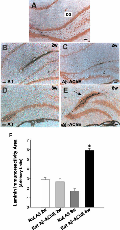
Figure 4.
Laminin immunoreactivity of amyloid deposits induced by Aβ-AChE complexes. A: Laminin is detected in the hippocampus associated to pyramidal neurons in the dentate gyrus in intact control animals. This immunoreactivity is focally diminished in the DG after 2 weeks of injection with Aβ fibrils (B) and with Aβ-AChE complexes (C). These results were maintained, after 8 weeks, in those animals injected with Aβ fibrils (D). However, in rats injected with Aβ-AChE complexes, the immunostaining was extensive (E) and it was concentrated extracellularly in the injection site (arrow), a fact that was not observed in the other groups. F: Laminin-immunoreactive area was quantified with the SigmaScan Pro software in adjacent brain sections. As observed in the graph, animals injected with Aβ-AChE complexes present a significant increase in laminin staining associated to the amyloid deposit in comparison to those injected with Aβ fibrils, 8 weeks after injection. *, P < 0.05. Scale bars, 100 μm.

