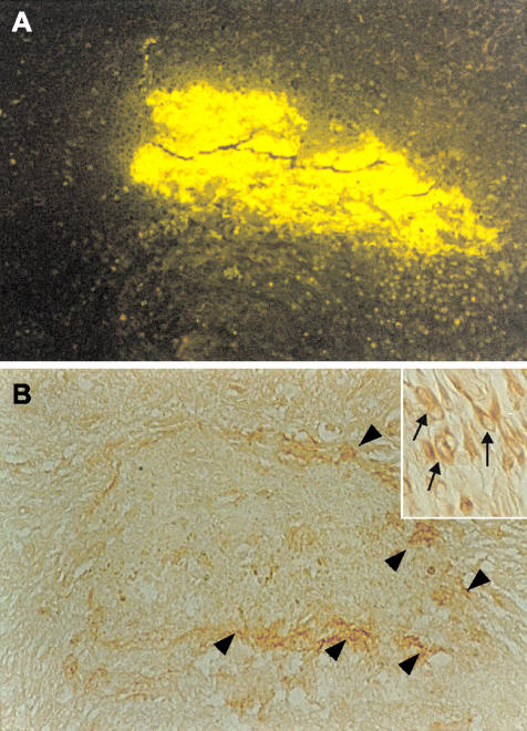
Figure 5.
Neurons that express laminin appear around the newly formed amyloid-AChE plaque. Animals were infused using an Alzet miniosmotic pump for 4 weeks with Aβ1-40 soluble peptide plus AChE into the hippocampus. A: Brain sections analyzed with Th-S present a big Th-S-positive deposit as a result of the infusion. B: Interestingly, adjacent sections showed laminin staining around the amyloid deposit (arrowheads) that correspond to neurons with increased laminin immunoreactivity (inset, arrows). Original magnifications: ×10; ×100 (inset).

