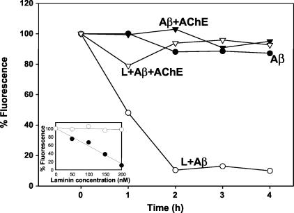
Figure 6.
Laminin disaggregates Aβ fibrils, but not Aβ-AChE complexes in vitro. Aβ1-40 fibrils (100 μmol/L) prepared either in the absence (circles) or the presence (inverted triangles) of AChE at a molar ratio of 1:1000 = AChE:Aβ were incubated either with 200 nmol/L of laminin (white symbols) or vehicle (black symbols). Th-T fluorescence (as expressed as percentage of respective time 0 points) of samples was taken at indicated time points. The inset shows the laminin concentration dependence of Th-T fluorescence formed in the presence (white circles) or the absence (black circles) of AChE at 2 hours of the assay.

