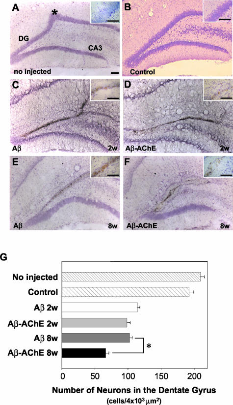
Figure 9.
Nissl staining (cresyl violet) at 2 and 8 weeks after Aβ fibril and Aβ-AChE complex injections. Nissl stained coronal sections through dorsal hippocampus near to the injection site (*) (intact animal A) in bilaterally injected rats with ACF+DMSO (control). DG indicates the dentate gyrus and CA3 indicates the region of the Ammon horn of the hippocampus (B), Aβ fibrils assembled alone after 2 (C) and 8 weeks (E), and Aβ-AChE complexes after 2 (D) and 8 weeks (F). Note the extensive neuronal degeneration in animals injected with Aβ-AChE complexes after 8 weeks. G: Pictures (×40) of Nissl-stained sections were submitted to quantification of the neuron number. As observed in the graph, only Aβ-AChE complexes after 8 weeks of treatment induced a significant neuronal cell loss in the upper leaf of the dentate gyrus. *, P > 0.05 (Student’s t-test). Scale bars, 100 μm.

