Figure 1.
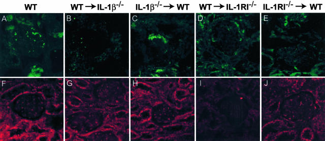
Renal localization of IL-β (green, A–E) and IL-1RI (red, F–J) in WT and chimeric mice with GN, detected by immunofluorescence, and captured by confocal microscopy. A: In glomeruli of WT mice, cytoplasmic staining of infiltrating leukocytes with occasional patchy staining of resident glomerular cells was observed. Tubular areas were also positive for IL-1β. B: In WT→IL-1β−/− chimeras IL-1β was only detected on macrophages infiltrating the kidney, no renal expression was detected. C: In IL-1β−/−→WT chimeric animals, IL-1β expression was sparsely observed in glomeruli and on tubular cells. In IL-1RI chimeric mice, WT→IL-1RI−/− (D), and IL-1RI−/−→WT (E), IL-1β expression was observed in a similar pattern to WT animals. Renal expression of IL-1RI in WT (F), WT→IL-1β−/− (G), and IL-1β−/−→WT (H) mice was similar, with the receptor observed to be present on cells in the glomerulus and on tubules. I: In contrast the WT→IL-1RI−/− chimeras had absent renal IL-1RI expression with limited expression detected on infiltrating inflammatory cells. J: The expression of IL-1RI in IL-1RI−/−→WT chimeras was not noticeably reduced compared with WT mice, this indicating that the prominent expression observed in WT and IL-1β chimeric mice to be by intrinsic renal cells. Confocal immunofluorescence, under oil immersion. Original magnifications, ×600.

