Figure 1.
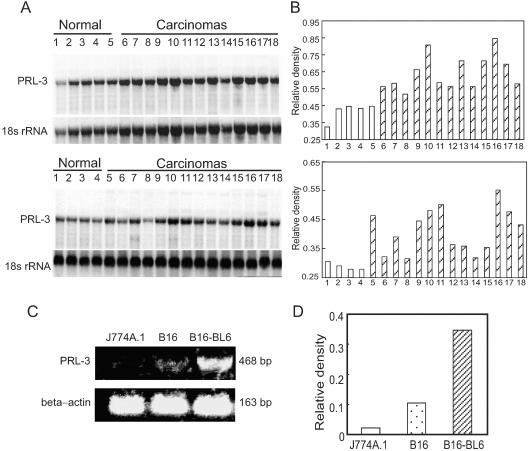
Expression of PRL-3 in liver carcinomas and tumor cell lines. A: Northern blot confirmed that the expression of the PRL-3 gene was enhanced in human liver carcinoma samples. Total RNA (10 μg/lane) extracted from the samples was used for analysis. The blot was sequentially probed to detect 18s rRNA. Top: Lanes 1 to 5, normal liver tissues; lanes 6 to 18, liver carcinoma samples. Bottom: Lanes 1 to 4, normal liver tissues; lanes 5 to 18, liver carcinoma samples. B: Histogram showing the PRL-3 transcript normalized to 18s rRNA. The values were presented as the percentage of 18s rRNA. C: Representative RT-PCR analysis for PRL-3 on mouse melanoma cell lines. The highly metastatic cell line B16-BL6 showed a higher level of mRNA encoding PRL-3 compared to the lowly metastatic cell line B16, as detected by RT-PCR. J774A.1 cells were PRL-3-negative. β-Actin was used as an internal control. D: The expression of PRL-3 mRNA comparing β-actin mRNA was quantified by densitometry. The values of transcript level for PRL-3 were normalized to β-actin transcript levels and were presented as the percentage of β-actin transcript.

