Figure 7.
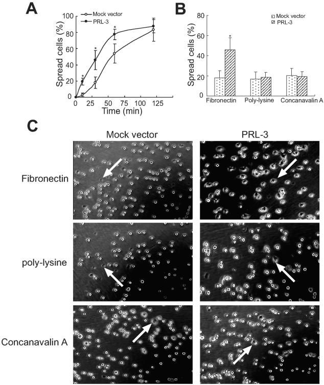
Enhanced cell spreading of PRL-3-transfected B16 cells. Cells were kept in suspension and then replated onto fibronectin-, polylysine-, or concanavalin A-coated cell culture dishes, incubated at 37°C, and then photographed at 10, 30, 60, and 120 minutes. A: Time course of cell spreading on fibronectin. B: Quantitative evaluation of cell spreading efficiency was obtained by calculating the percentage of cells spread on fibronectin, polylysine, or concanavalin A at 30 minutes. Shown are mean percentages of spread cells per field ± SEM of three independent experiments. At least 10 different fields for each sample were averaged. A total of ∼500 cells were counted for each bar. *, P < 0.05, versus mock vector. C: Representative photographs from mock vector- and PRL-3-transfected cells after spread for 30 minutes. The arrows indicated the spreading cells with flat shape and dark phase.

