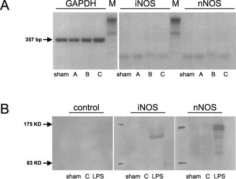
Figure 1.
A: Detection of GAPDH (357 bp), iNOS (468 bp) and nNOS transcripts (281 bp) by RT-PCR in RNA extracted from sham-operated lungs (sham) and left lungs exposed to 1 hour of ischemia, followed by either 0.5 hours of reperfusion (group A), 5 hours of reperfusion (group B), or 24 hours of reperfusion (group C). Experiments were performed in WT animals. B: Western blot analysis of iNOS and nNOS protein in sham-operated control lung tissue (sham) and in lung tissue exposed to 1 hour of ischemia followed by 24 hours of reperfusion (group C). Protein from lung tissue (iNOS) and brain tissue (nNOS) harvested from LPS-exposed animals served as positive control (LPS). To assess unspecific background, controls without primary antibody were performed. Experiments were performed in WT animals.

