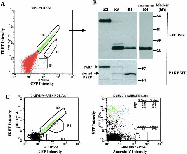
Figure 3.
Analysis of PARP cleavage and annexin V binding in cells transfected with the CFP-LEVD-YFP probe. A: Flow cytometric profile showing a population with bright FRET (R2, intact CFP-LEVD-YFP molecules) and a population with diminished FRET that could be further classified into R3 (incomplete cleavage of the probe), and R4 (complete cleavage of the probe). B: The GFP and PARP immunoblots confirmed the cleavage of the probe and of the endogenous caspase-sensitive substrate PARP in the regions indicated in (A). C: Apoptosis revealed by annexin V staining demonstrated annexin V binding by the population with diminished FRET. Hela cells transfected with the caspase-sensitive probe were stained with APC-conjugated annexin V and then were analyzed for FRET and annexin V binding. Three different regions as indicated in (A) in a plot of CFP versus FRET intensity were gated and further visualized in a plot of annexin V versus YFP intensity. The blue cells in R4, but not those in R2, bound annexin V weakly. Spontaneous caspase activity was documented in 1.2 percentages of all cultured Hela cells (R5).

