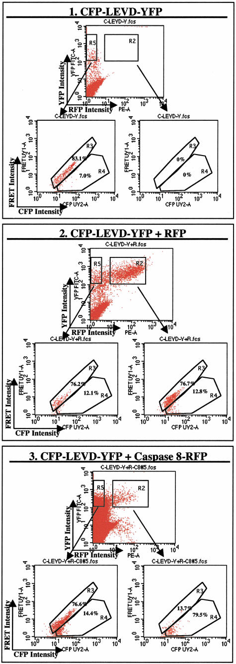
Figure 6.
The caspase-sensitive CFP-LEVD-YFP probe is cleaved in Hela cells overexpressing human caspase 8. A fusion protein consisting of human caspase 8 and red fluorescent protein (RFP) was used for these experiments. With the four-color two-laser optical configuration shown in Figure 1A, CFP, FRET, YFP, and RFP signals could be easily distinguished. For these experiments, the red fluorescent protein signal was assessed with the FL2 detector. Cells were transfected with the caspase-sensitive CFP-LEVD-YFP probe with or without caspase 8-RFP or RFP. Transfected cells expressing YFP (and CFP) along with RFP are indicated in R2, whereas the cells expressing YFP (and CFP) but not RFP are gated in R5. FRET was then assessed for each population and the FRET-positive cells and cells with diminished FRET in R2 and R5 were analyzed. (1). Flow cytometric profiles showing cells transfected with the caspase-sensitive CFP-LEVD-YFP probe only. There were nearly no events gated in R2. The cells within R5 had a similar pattern in a plot of CFP versus FRET signals as shown in Figure 2 with a large population of FRET-positive cells and fewer cells with diminished FRET. (2). Flow cytometric profiles showing cells co-transfected with the caspase-sensitive CFP-LEVD-YFP probe and RFP. The patterns of FRET-positive and cells with diminished FRET in R2 and R5 were similar. (3). Flow cytometric profiles showing that caspase 8 cleaved the caspase-sensitive CFP-LEVD-YFP probe in vivo. The cells expressing caspase 8-RFP (R2) manifested a markedly increased fraction of cells with diminished FRET (79.5%) compared to cells in R5 that did not express caspase 8-RFP (14.4%) or in cells transfected with CFP-LEVD-YFP alone (7.0%) or cells transfected with CFP-LEVD-YFP plus RFP (12.1 to 12.8%).

