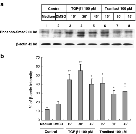
Figure 5.
Activation of Smad2 by Tranilast. (a) HaCaT cells were treated with 100 pmol l−1 (pM) TGF-β1 (lanes 3, 4 and 5) or 100 μmol l−1 (μM) Tranilast (lanes 6, 7 and 8). The cells were harvested every 15 min for protein analysis. Total proteins from cell lysates were separated and probed with anti-phospho-Smad2 antibody. Phospho-Smad2 is a 60-kDa protein. The β-actin was also probed to demonstrate the equal loading of protein for each group. (b) Quantitative analysis with β-actin normalization showed significant stimulatory effects of TGF-β1 and Tranilast on protein expression level of phospho-Smad2. Each experiment was repeated three times. *P<0.05; **P<0.01 as compared to medium control in each group.

